
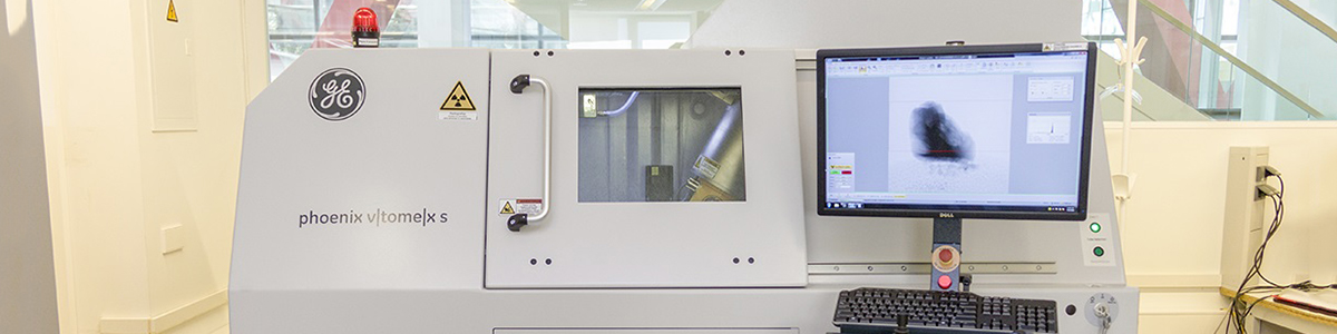
Micro-Computed Tomography
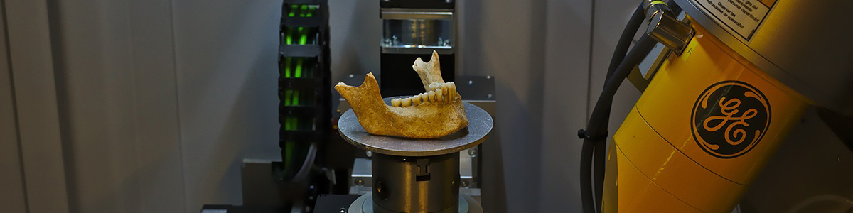
This laboratory boasts a microCT (model V|Tome|X s 240 by GE Sensing & Inspections Technologies) along with the corresponding reconstruction and image analysis softwares. The variation of the parameters of the x-ray tube allow us to select the best set-up depending on the object under analysis, whether rock, bone, polymer, concrete, teeth, tissue, etc. The equipment can scan samples with a weight of up to 10 kg, a height of up to 40 cm and a diameter of up to 20 cm.
The laboratory is part of the CENIEH's outstanding facilities, so it can be accessed through Competitive Access calls through the User Office.
Micro-Computed Tomography is based on the interaction between the radiation and the material (according to the Beer–Lambert law). In other words, the radiation transmitted is the information captured by a detection system in image form (radiography). In this manner, a "slice" of material within a solid can be described, based on the different images taken at various different angles. During the tomographic reconstruction process, the main objective is to reconstruct an object that is considered as a 2D distribution of some type of function μ(x,y), which represents the attenuation coefficient of the object in question. The reconstruction of the object is obtained by resolving a mathematical problem, generally implemented in several packages of software.
This reconstruction process makes it possible to easily obtain a volumetric representation of the object, enabling the study and evaluation of different parameters thereof (for example: dispersion of particles, detection of defects, density profiles, etc.).
It can therefore be stated that micro-computed tomography is an x-ray image technique that makes it possible to scan, explore and model samples in 3D. Thus, it is a NON-destructive technique, the basic principle of which is the virtual reconstruction of the sections of an object based on images (radiographies) that have been acquired over 360º.
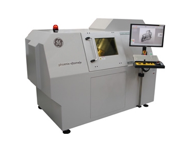
MicroCT V|Tome|X s 240 by GE Sensing & Inspections Technologies Phoenix X-Ray: A versatile x-ray inspection system for Micro-Computed Tomography (Micro-CT and Nano-CT). The maximum diameter and length of the sample to be scanned in 3D is 200 mm x 400 mm, with a maximum weight of 10 kg. It is highly versatile thanks to the two x-ray tubes by which it is formed: the 240 KV microfocus with detectability values of 1 µm, and the 180 KV nanofocus, with detectability values below 0.5 µm.
TESCAN CoreTOM MicroCT:
This equipment has been funded by the European Union – NextGenerationEU within the framework of the Recovery, Transformation and Resilience Plan.
Versatile X-ray inspection system for computed tomography, equipped with a microfocus tube (maximum resolution of 2–3 microns; maximum power of 300 W). The maximum sample size that can be scanned is 60 cm in diameter and 1 m in height, with a weight limit of 45 kg. The system is specially designed for in-situ testing and currently includes the necessary module for performing mechanical tensile and compression tests.
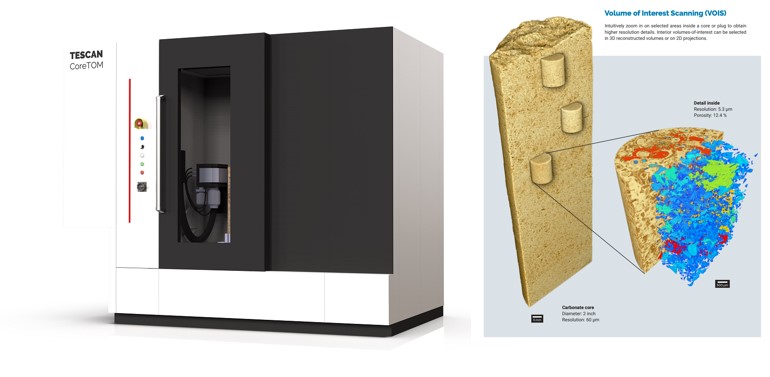
CoreTOM VOIS-CoreTOM
AVIZO: Software developed by Thermo Fisher for processing images from computed microtomography or microscopy, enabling the analysis of these images for advanced material characterization and 3D visualization.
DragonFly ORS: Software developed by ORS for processing images from computed microtomography or microscopy, enabling the analysis of these images for advanced material characterization and 3D visualization.
Applications
- Materials science (detection of defects, porosity, connectivity of pores, detection of different phases/materials, density profiles, etc.)
- Quality control
- Medicine
- Biology
Services
- Collection of results in parent files: TIFF, DICONDE, RAW, etc.
- 3D reconstruction of files from other analyses that have already been created
- Customer use of the software Mimics 16.0 (Materialise) for the image analysis of samples
- Advice in terms of image analysis
- Digital image analysis
- Users of the Microcomputed Tomography service who are going to scan cultural heritage items. The Conservation and Restoration lab offer the option of complementing it, at no additional cost, in order to preparing the scan.
- The temporary storage of their cultural assets will be made available to users in the CENIEH collection rooms provided at the Conservation and Restoration Laboratory. The storage will be done only in case that the nature of the cultural assets are compatible with the conservation conditions established in this collections rooms

Microscopy and Micro-Computed Tomography Laboratory Technician